 |
|

 Shoulder
instability develops in two different ways: traumatic
onset (related to a sudden injury) or atraumatic onset
(not related to a sudden injury). Understanding the differences is
essential in choosing the best course of treatment. As a rule, the
patient with atraumatic onset instability has general
laxity (looseness) in the joint that eventually causes
the shoulder to become unstable, whereas traumatic
onset instability begins when an injury causes a shoulder to develop
recurrent (repeated) dislocations. Shoulder
instability develops in two different ways: traumatic
onset (related to a sudden injury) or atraumatic onset
(not related to a sudden injury). Understanding the differences is
essential in choosing the best course of treatment. As a rule, the
patient with atraumatic onset instability has general
laxity (looseness) in the joint that eventually causes
the shoulder to become unstable, whereas traumatic
onset instability begins when an injury causes a shoulder to develop
recurrent (repeated) dislocations.
Atraumatic shoulder instability, also called multidirectional instability
(MDI), is described as laxity of the shoulder's glenohumeral joint
in multiple directions.
What
does the inside of the shoulder look like?
The shoulder is the most mobile joint in the human body with a complex
arrangement of structures working together to provide the movement
necessary for daily life. Unfortunately, this great mobility comes
at the expense of stability. Four bones and a network of soft
tissues (ligaments, tendons, and muscles), work together to
produce shoulder movement. They interact to keep the joint in place
while it moves through extreme ranges of motion. Each of these structures
makes an important contribution to shoulder movement and stability.
Certain work or sports activities can put great demands upon the shoulder,
and injury can occur when the limits of movement are exceeded and/or
the individual structures are overloaded. Click
here to read more about shoulder structure.
What is atraumatic shoulder instability?
 Atraumatic shoulder instability develops in patients who have increased
looseness of the supporting ligaments that surround the shoulder's
glenohumeral joint. The laxity can be a natural condition (present
from birth) or a condition that has developed over time. Many patients
with MDI are active in overhead sports (such as gymnastics, swimming,
or throwing) that repetitively stretch the shoulder capsule to extreme
ranges of motion. Atraumatic shoulder instability develops in patients who have increased
looseness of the supporting ligaments that surround the shoulder's
glenohumeral joint. The laxity can be a natural condition (present
from birth) or a condition that has developed over time. Many patients
with MDI are active in overhead sports (such as gymnastics, swimming,
or throwing) that repetitively stretch the shoulder capsule to extreme
ranges of motion.
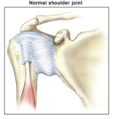
The glenoid (the socket of the shoulder joint) is a
relatively flat surface that is deepened slightly by the labrum,
a cartilage cup that surrounds part of the head of the humerus. The
labrum acts as a bumper to keep the humeral head firmly in place in
the glenoid. It is also the attachment point for important ligaments
that stabilize the shoulder. These ligaments often become stretched
out with MDI, allowing dislocation or subluxation (an incomplete
or partial dislocation) to occur. The increased motion of the
joint can lead to repetitive microtrauma (small injuries),
producing tears of the labrum or rotator cuff.
MDI patients will often have increased ligament laxity in many joints.
Hyperextended knees, elbows, and a self-described history of being
"double-jointed" are common. These patients often have multidirectional
laxity in both shoulders. Because many athletes with MDI are quite
successful in their sports, there is a debate about whether laxity
improves performance or is caused by repetitive stretching during
athletic activity.
|

© 2015 by LeadingMD.com All rights reserved.
Disclaimer
|
| |
|
Stem cells, PRP, and HA, oh my! We are not in Kansas anymore… Part 2
READ MORE >>
|
|
|
|

