|

The Surgical Procedure: Microfracture
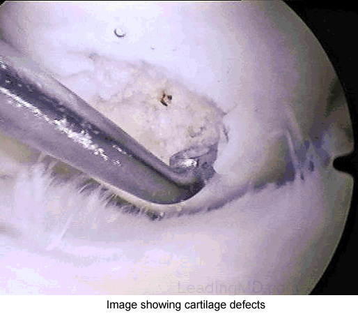
The microfracture procedure is done arthroscopically.
The surgeon visually assesses the defect and performs the procedure
using special instruments that are inserted through three small
incisions on the knee. After assessing the cartilage damage, any
unstable cartilage is removed from the exposed bone. The surrounding
rim of remaining articular cartilage is also checked for loose or
marginally attached cartilage. This loose cartilage is also removed
so that there is a stable edge of cartilage surrounding the defect.
The process of thoroughly cleaning and preparing the defect is
essential for optimum results.
Multiple holes, or microfractures, are then made in
the exposed bone about 3 to 4mm apart. Bone marrow cells and
blood from the holes combine to form a "super clot" that
completely covers the damaged area. This marrow-rich clot is
the basis for the new tissue formation. The microfracture technique
produces a rough bone surface that the clot adheres to more easily.
This clot eventually matures into firm repair tissue that becomes
smooth and durable. Since this maturing process is gradual, it usually
takes two to six months after the procedure for the patient to experience
improvement in the pain and function of the knee. Improvement is
likely to continue for about 2 to 3 years.
What types of complications may occur?
Most patients progress through
the postoperative period with little or no difficulty. Some patients
may develop mild transient pain, most frequently after microfracture
has been performed on the patella (kneecap) and trochlear
groove (the groove on the femur in which the patella glides
during motion). Small changes in the articular surface of this patellofemoral
joint may produce a grating or "gritty" sensation, particularly
when a patient discontinues use of the knee brace and begins normal
weightbearing through a full range of motion. Patients rarely have
pain at this time, and this grating sensation typically resolves
on its own in a few days or weeks.
Similarly, if a steep
perpendicular rim was made in the trochlear groove, patients may
notice "catching" or "locking" as the ridge
of the patella rides over this area during joint motion. Some patients
may even notice these symptoms while using the continuous
passive motion machine (CPM), a device that gently moves
the joint while the patient is lying down. If this locking sensation
is painful, the patient is advised to limit weightbearing and avoid
the bothersome joint angle for an additional period. These symptoms
usually dissipate within 3 months.
Typically, swelling and joint effusion
(fluid in the joint) disappear within 8 weeks after a microfracture
procedure. Occasionally, a recurrent effusion develops between 6
and 8 weeks after surgery for a defect on the femur; usually when
a patient begins to put weight on the injured leg. This effusion
may mimic the preoperative or immediate postoperative effusion,
although it is usually painless. It usually resolves within several
weeks. Rarely is a second arthroscopy required for recurring effusions.
|